
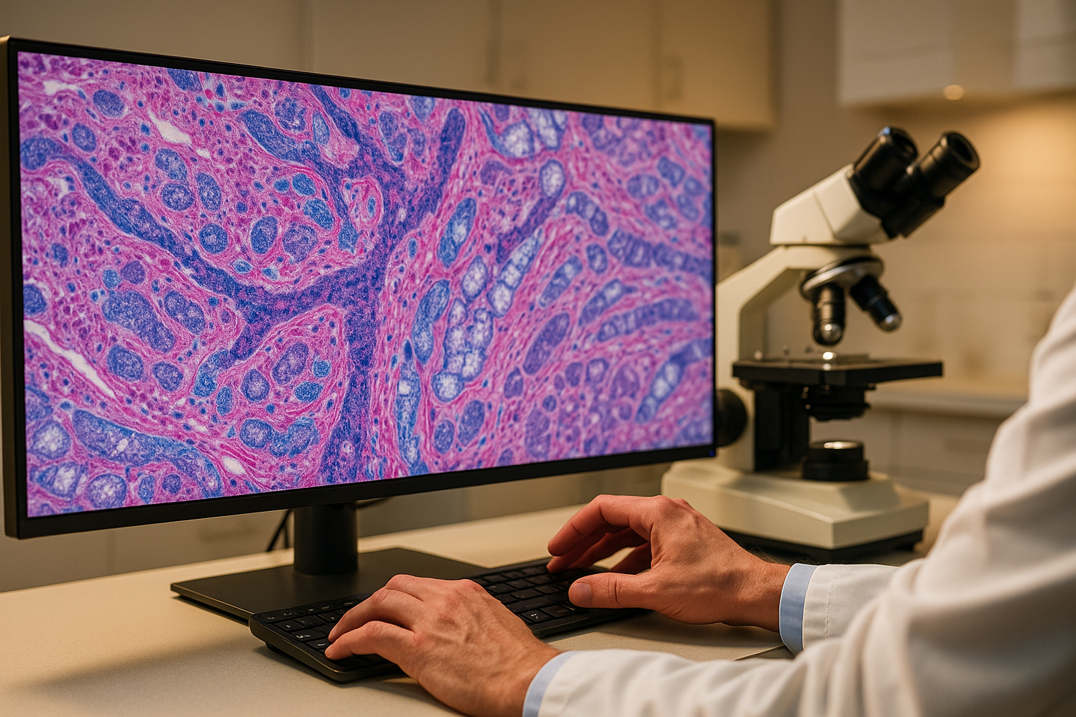
Digital pathology is undergoing a revolution. The marriage of whole slide imaging (WSI) with artificial intelligence is creating systems capable of detecting cancers, grading tumors, predicting molecular markers, and identifying patterns invisible to the human eye. At the heart of this revolution are open source datasets — massive collections of digitized tissue slides with expert annotations that enable researchers worldwide to develop and validate computational pathology algorithms.
This guide provides an exhaustive catalog of every major open source pathology dataset available for AI research, with direct links to download portals and code repositories. Whether you are working on cancer detection, cell segmentation, survival prediction, or foundation model development, this is your complete reference.
The Digital Pathology Data Landscape
Pathology datasets present unique computational challenges. A single whole slide image (WSI) can be 100,000 x 100,000 pixels or larger — gigapixel-scale images that cannot be processed by standard deep learning pipelines without patch-based or multiple instance learning approaches. This scale, combined with the complexity of tissue morphology, makes pathology one of the most technically demanding areas of medical imaging AI.
Datasets in this space range from small, meticulously annotated collections of a few hundred slides to massive multi-institutional archives containing tens of thousands of cases. The annotations themselves vary from coarse slide-level labels to pixel-level segmentations of individual cells and tissue structures.
Large-Scale Whole Slide Image Archives
These repositories serve as foundational resources, providing access to thousands of digitized slides across multiple cancer types.
| Dataset | Slides | Cancer Types | Annotations | Source |
|---|---|---|---|---|
| TCGA (The Cancer Genome Atlas) | 30,000+ diagnostic slides | 33 cancer types | Slide-level diagnoses; paired genomics, transcriptomics, clinical data | GDC Portal |
| CPTAC (Clinical Proteomic Tumor Analysis) | 3,500+ slides | 10+ cancer types | Paired proteomics and histopathology | NCI CPTAC |
| GTEx | 17,382 tissue samples | Normal tissues (54 sites) | Gene expression paired with histology | gtexportal.org |
| PAIP (Pathology AI Platform) | Varies by challenge year | Liver, colon, pancreas | Tumor segmentation and viable tumor burden estimation | Grand Challenge |
Breast Cancer Pathology Datasets
Breast cancer pathology has the richest ecosystem of open datasets, reflecting its clinical importance and the maturity of AI research in this domain.
| Dataset | Size | Task | Annotations | Source |
|---|---|---|---|---|
| Camelyon16 | 399 WSIs | Lymph node metastasis detection | Pixel-level metastasis annotations; landmark challenge in pathology AI | Grand Challenge |
| Camelyon17 | 1,000 WSIs from 5 centers | Patient-level pN-staging | Multi-center extension with clinical staging | Grand Challenge |
| BreakHis | 9,109 microscopy images | Benign vs. malignant classification | 4 magnification levels (40x, 100x, 200x, 400x); 8 tumor subtypes | UFPR |
| BACH (BreAst Cancer Histology) | 400 images + 30 WSIs | 4-class classification | Normal, benign, in situ carcinoma, invasive carcinoma | Grand Challenge |
| BRACS | 4,539 ROIs + 547 WSIs | 7-class breast lesion classification | Atypical ductal hyperplasia included; region and WSI-level annotations | BRACS |
| TUPAC16 | 821 WSIs | Mitosis detection + proliferation scoring | Mitosis annotations; tumor proliferation speed prediction | Grand Challenge |
Colorectal and Gastrointestinal Pathology
| Dataset | Size | Task | Annotations | Source |
|---|---|---|---|---|
| NCT-CRC-HE-100K | 100,000 patches | 9-class tissue classification | Adipose, background, debris, lymphocytes, mucus, smooth muscle, normal mucosa, cancer stroma, tumor epithelium | Zenodo |
| GlaS (Gland Segmentation) | 165 images | Gland instance segmentation | Pixel-level gland boundaries in colon adenocarcinoma | TIA Warwick |
| DigestPath 2019 | 872 images | Signet ring cell detection + colonoscopy tissue segmentation | Cell-level and tissue-level annotations | Grand Challenge |
| CRAG | 213 images | Colorectal adenocarcinoma gland segmentation | Instance-level gland segmentation with grading | TIA Warwick |
Cell Detection and Segmentation Datasets
Cell-level analysis is fundamental to computational pathology, enabling quantification of tumor-infiltrating lymphocytes, mitotic counts, and cellular composition.
| Dataset | Size | Task | Annotations | Source |
|---|---|---|---|---|
| PanNuke | 7,901 patches from 19 tissue types | Nuclei instance segmentation | 5 nuclei types: neoplastic, inflammatory, connective, dead, epithelial | TIA Warwick |
| CoNSeP | 41 H&E image tiles | Nuclei segmentation and classification | 24,319 annotated nuclei with 7 cell types | TIA Warwick |
| MoNuSeg | 44 H&E images from 7 organs | Nuclear segmentation | 21,623 manually annotated nuclear boundaries | Grand Challenge |
| Lizard | 291 H&E patches | Nuclear instance segmentation | 495,179 nuclei labeled into 6 classes across colon tissue | TIA Warwick |
| NuCLS | 1,744 ROIs from breast cancer | Nuclear classification | 220,000+ nuclei with 13 cell type labels | NuCLS |
Prostate and Kidney Pathology
| Dataset | Size | Task | Annotations | Source |
|---|---|---|---|---|
| PANDA (Prostate Cancer Grade Assessment) | 10,616 WSIs | Gleason grading | Slide-level ISUP grades from 2 European centers; largest prostate pathology dataset | Kaggle |
| SICAPv2 | 18,783 patches from 155 WSIs | Gleason pattern classification | Patch-level Gleason pattern (3, 4, 5) and non-cancerous annotations | Mendeley Data |
| HuBMAP Kidney | 20 WSIs | Functional tissue unit segmentation | Glomeruli and tubule segmentation in PAS-stained kidney | Kaggle |
Foundation Models and Pretrained Weights
The pathology AI community has produced several open source foundation models that can be fine-tuned on smaller datasets:
| Model | Architecture | Training Data | Key Features | Source |
|---|---|---|---|---|
| UNI | ViT-Large | 100M+ patches from 100K+ WSIs | General-purpose pathology feature extractor; state-of-the-art on 34 benchmarks | GitHub |
| CONCH | Vision-Language | 1.17M image-text pairs | Contrastive learning from pathology images and captions | GitHub |
| CTransPath | Swin Transformer | 15M patches from 30K+ slides | Semantically relevant contrastive learning for histopathology | GitHub |
| Phikon | ViT-Base | TCGA + CPTAC slides | Self-supervised pathology model from Owkin | Hugging Face |
| HoverNet | Custom encoder-decoder | PanNuke + CoNSeP | Simultaneous nuclei segmentation and classification | GitHub |
Tools and Frameworks
Several open source tools make working with these datasets more accessible:
- CLAM — Data-efficient and weakly supervised computational pathology pipeline
- MITI Minimum Information Standard — Standardized reporting for tissue image datasets
- QuPath — Open source whole slide image analysis software
- deep-histopath — Deep learning framework for computational pathology
- MONAI Pathology — Pathology-specific extensions to the MONAI framework
Getting Started
For researchers entering computational pathology, we recommend starting with NCT-CRC-HE-100K for tissue classification (patch-level, manageable size) or Camelyon16 for WSI-level analysis. The PANDA dataset is excellent for learning multiple instance learning approaches on a well-documented, large-scale task. For cell-level analysis, PanNuke provides the breadth needed across tissue types.
The field is moving rapidly toward foundation models, and researchers should consider leveraging pretrained weights from UNI, CONCH, or Phikon rather than training from scratch — particularly for smaller downstream datasets.
As computational pathology matures, we anticipate more datasets incorporating immunohistochemistry, special stains, and spatial transcriptomics data, enabling multimodal models that capture the full complexity of tissue biology.