Melanoma accounts for a small fraction of skin cancer cases but a disproportionate share of skin cancer deaths, largely because outcomes depend heavily on catching disease early. Dermatology has long relied on visual pattern recognition—making it a natural fit for machine learning (ML) systems trained to detect malignancy from images. Over the past decade, AI for melanoma detection has matured from research prototypes into a growing ecosystem of tools that support triage, documentation, and clinical decision-making. Still, the best results come not from “AI replacing dermatologists,” but from careful integration into workflows—paired with rigorous validation, bias testing, and clear guardrails for patient safety.
Why dermatology is an early proving ground for clinical AI
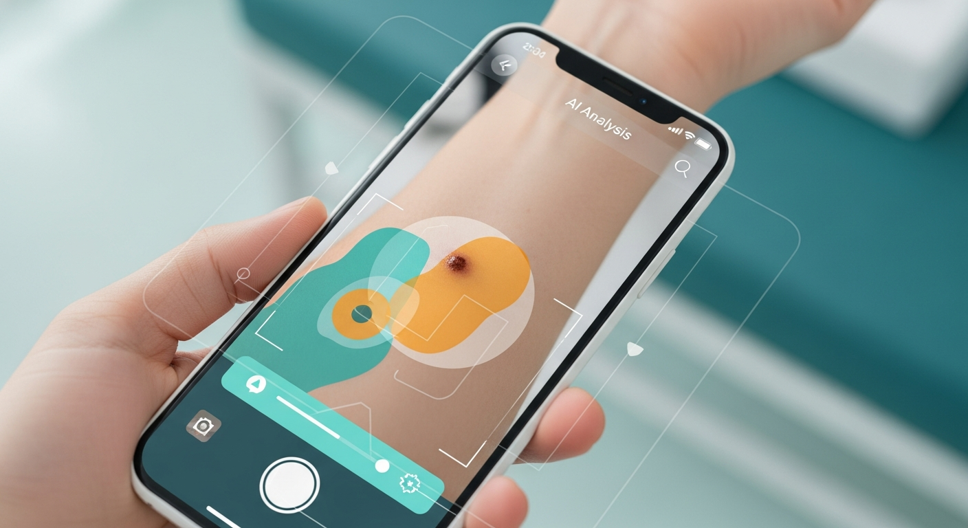
Dermatology is image-rich and comparatively standardized: lesions can be photographed with consumer smartphones, dermatoscopes, or high-resolution clinical cameras. That makes it possible to build large labeled datasets for supervised learning and to evaluate model performance in controlled test sets. In practice, melanoma detection AI tends to fall into three overlapping categories:
- Consumer-facing risk assessment: smartphone apps and camera-based tools that estimate whether a mole looks suspicious.
- Clinical decision support: AI that helps clinicians triage lesions, prioritize referrals, or support biopsy decisions.
- Workflow and documentation: tools that standardize imaging, track lesions over time, and integrate with the EHR.
Most melanoma-focused AI systems use deep convolutional neural networks (CNNs) or vision transformers trained on dermoscopic images, clinical photos, or both. Performance is usually reported using metrics like sensitivity (catching true melanomas), specificity (avoiding false alarms), and area under the ROC curve (AUC).
What the research says: strong benchmarks, messy real-world deployment
Academic momentum accelerated after widely cited work showed that deep learning models could match or exceed dermatologist-level performance on curated image sets. A landmark example is the 2017 Nature paper by Esteva and colleagues, which trained a CNN on a large dataset of skin lesion images and reported performance comparable to dermatologists on benchmark tasks (Nature, 2017). That study helped set the narrative—but it also highlighted a recurring challenge: models can look excellent on benchmark datasets yet stumble when exposed to the variability of real-world practice.
More recent research has focused on generalizability (does the model work across different devices, lighting, and clinical sites?), fairness (does performance hold across skin tones and demographic groups?), and prospective validation (does it improve outcomes in real clinical workflows?). Researchers have also explored hybrid approaches that combine dermoscopic images with clinical metadata (age, lesion location, personal history), which can improve discrimination but complicates deployment because structured data quality varies across settings.
Key technical and clinical friction points
- Dataset shift: Training images are often captured under ideal conditions; real images include blur, glare, occlusion, and inconsistent framing.
- Label noise: Even biopsy-confirmed labels have nuance (atypical nevi, borderline lesions), while many datasets rely on clinician assessment rather than histopathology.
- Skin tone representation: Underrepresentation of darker skin in dermatology datasets can degrade accuracy and increase missed diagnoses in groups already facing disparities.
- Clinical thresholds: A “good” AUC can still be unsafe if the chosen operating point misses melanoma or generates unmanageable false positives.
Google Lens and the consumerization of visual search
When people find something unfamiliar on their skin, many now start with a smartphone. While Google Lens is not a regulated medical device and is marketed as a general-purpose visual search tool, it has become part of the de facto consumer health pathway: users take photos and search for visually similar images. This matters clinically for two reasons. First, it can influence patient anxiety, self-triage, and timing of care-seeking. Second, it underscores a broader trend: image-based AI is increasingly ambient—embedded in everyday tools rather than limited to clinical software.
From a safety perspective, general-purpose image search is not the same as clinical AI: it may return look-alike images without calibrated risk estimates, clinical context, or guidance on urgency. Dermatology clinics are already seeing the downstream effect—patients arrive with screenshots and strong expectations. The opportunity for clinical AI is to provide a safer bridge: validated tools that can prompt urgent evaluation for high-risk lesions, while discouraging false reassurance.
Notable projects and new directions in melanoma AI
Melanoma detection has moved beyond “single-image classification” toward systems that better reflect clinical practice.
1) Multi-modal and longitudinal models
Newer projects aim to combine dermoscopy with standard clinical photos and patient metadata, and to track lesions over time. Longitudinal comparison—detecting change in size, color, or border irregularity—mirrors how dermatologists monitor atypical moles and can reduce unnecessary biopsies. This also aligns with the growing interest in foundation models for medical imaging, which can be fine-tuned for specific tasks like pigmented lesion classification.
2) Prospective and workflow-integrated evaluation
The field is increasingly emphasizing prospective studies and clinic-based pilots over retrospective benchmark performance. These evaluations ask practical questions: Does AI reduce time-to-biopsy for true melanomas? Does it change clinician decision-making? Does it overload clinics with false positives? And how does it perform on diverse populations and devices?
3) Dermoscopy quality control and “human-in-the-loop” design
A quiet but important innovation is AI that checks image quality (focus, illumination, framing) before analysis. Another is decision support that explains model attention (e.g., saliency maps) and communicates uncertainty. In many clinics, the safest pattern is a human-in-the-loop approach: AI flags concerning lesions and supports documentation, while clinicians retain diagnostic responsibility and determine whether biopsy is warranted.
Clinical impact: where AI helps today
When deployed responsibly, melanoma AI can deliver measurable benefits:
- Triage support: prioritizing high-risk referrals and reducing time-to-specialist for suspicious lesions.
- Decision support: helping clinicians—especially non-dermatologists—decide when to refer or biopsy.
- Access and scalability: supporting teledermatology by standardizing image capture and pre-screening large volumes of cases.
- Consistency: reducing variability in assessments between clinicians and across sites.
These gains are especially relevant in primary care and underserved areas where dermatology shortages can delay evaluation.
Safety, regulation, and the risk of overconfidence
Melanoma is a high-stakes target: the cost of a false negative can be life-threatening, while excessive false positives can drive unnecessary biopsies, scarring, anxiety, and system burden. For publication-grade and clinical-grade AI, the most important questions are not just “How accurate is it?” but:
- Validated on what population? Including a representative range of skin tones, ages, and lesion types.
- Validated in what setting? Dermoscopy images from specialty clinics may not match smartphone photos from primary care.
- What’s the intended use? Consumer triage vs. clinician decision support require different thresholds and messaging.
- How is uncertainty handled? Systems should fail safely, prompting clinical evaluation when confidence is low.
Regulators have increasingly emphasized transparency around intended use, performance evidence, and post-market monitoring for AI/ML-based software. For healthcare organizations, governance also includes model monitoring (detecting performance drift), cybersecurity protections for image data, and clear patient communication that AI is assistive—not definitive.
What to watch next
Three developments will shape the next phase of melanoma detection AI:
- Foundation models in dermatology: larger pre-trained vision models fine-tuned for lesion analysis, potentially improving robustness across devices and settings.
- Better equity benchmarks: standardized reporting across Fitzpatrick skin types and demographic groups, moving fairness from a footnote to a requirement.
- Integrated care pathways: AI that links detection to action—streamlined referral, telederm consult, and follow-up—rather than standalone “risk scores.”
In parallel, consumer tools like Google Lens will continue to influence how patients interpret skin changes. That makes it even more important for clinical AI developers—and healthcare systems—to provide validated, context-aware alternatives that encourage timely care without amplifying misinformation or false reassurance.
Bottom line
AI for melanoma detection is one of the most promising and visible applications of clinical computer vision. The science has advanced well beyond proof-of-concept, with strong benchmark performance and an expanding range of real-world pilots. The next leap will depend on prospective evidence, equitable performance across skin tones, and practical integration into care pathways—so AI improves outcomes, not just accuracy charts.
References (selected): Esteva A. et al., “Dermatologist-level classification of skin cancer with deep neural networks,” Nature (2017). Google Lens (Google) as a general-purpose visual search product frequently used by consumers for image-based queries; not a regulated diagnostic tool.